Davies, AJ, Chaplin, TA, Rosa, MJP & Yu 2016, ‘Natural motion trajectory enhances the coding of speed in primate extrastriate cortex’. Scientific Reports, 6: 19739.
Associated Institution(s): Monash University and Australian Research Council Centre of Excellence for Integrative Brain Function
The Experiments
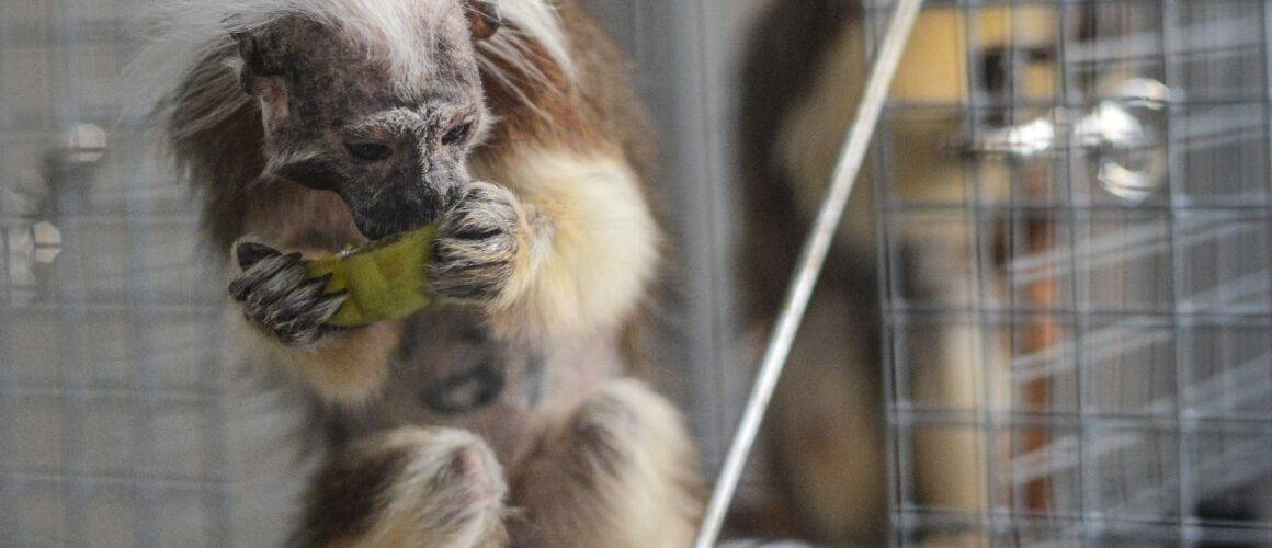
Six adult marmosets were used for visual mapping experiments.
Visual motion is believed to be processed by neurons in the middle temporal area of the human brain. In these experiments recordings were obtained from either the primary visual cortex and/or the middle temporal area of the brains of marmosets.
In order to prepare the monkeys for the experiment they each had to undergo the following procedure developed in 2003 by Bourne and Rosa(1). It should be noted that these procedures are carried out not only in this experiment but are used as a standard procedure in many experiments where animals have to be restrained for brain research.
Animals have food withdrawn for 12 hours prior to anaesthesia. A tracheotomy (an incision in the windpipe) is firstly performed which is then followed by cannulation of the femoral vein in the thigh and catheterisation of the urinary tract.
The monkeys are then placed in a stereotaxic frame fitted with jacks (covered with a homeothermic blanket and layers of absorbent pads) used to raise the animals to the correct level and to hold them completely still. Needles are inserted between the chest wall and forelimbs and connected to a monitor.
Next a craniotomy is performed (surgical removal of part of the skull to expose the brain). Once the cortex is exposed an acrylic well is constructed around the craniotomy. Small burr holes are drilled into the skull, filled with dental acrylic which forms a base for a stainless steel bolt affixed to the skull and secured onto the stereotaxic frame. The eyes are held open and covered with contact lenses. Whilst chemically paralysed and artificially ventilated the brain recordings take place.
Relevance to Humans
There are major anatomical, genetic, dietetic, environmental, toxic, and immune differences between animals – including marmosets – and humans(2), making them inappropriate for use in studying human brain injury and human disease. Many studies and systematic reviews show that there is discordance between animal and human studies, and that animal ‘models’ fail to mimic clinical disease adequately.(3)(4)
Funding
The experiment was funded through National Health and Medical Research Council of Australia (NHMRC) project grant number 1020839 – $548,938, project grant number 1083152 – $624,514) and Australian Research Council (DE1411505, CE140100007).
References
(1) Bourne, JA & Rosa, MGP 2003, ‘Preparation for the in vivo recording of neuronal responses in the visual cortex of anaesthetised marmosets (Callithrix jacchus)’, Brain Research Protocols, 11: 168–177.
(2) Pound P, Ebrahim S, Sandercock P, Bracken MB, Roberts I; on behalf of the Reviewing Animal Trials Systematically Group. 2004. ‘Where is the evidence that animal research benefits humans?’, BMJ, 328, 514-7.
(3) Perel P, Roberts I, Sena E, Wheble P, Briscoe C, Sandercock P, et al. 2007. ‘Comparison of treatment effects between animal experiments and clinical trials: systemic review’, BMJ, 334:197.
(4) Van der Worp H, Howells DV, Sena ES, Porritt MJ, Rewell S, O”Collins V, et al. 2010. ‘Can animal models of disease reliably inform human studies?’, PLoS Med.
